
New Study Identifies Two Distinct Gray Matter Patterns in Autism Spectrum Disorder

Understanding Neuroanatomical Diversity in Autism
Autism spectrum disorder (ASD) is known for its broad range of symptoms and brain differences, making it challenging to pinpoint consistent neuroanatomical features. While previous magnetic resonance imaging (MRI) studies have reported both increases and decreases in gray matter volume compared to typically developing individuals, these mixed findings may mask distinct brain patterns within the autism population.
Applying Advanced Subtyping Techniques
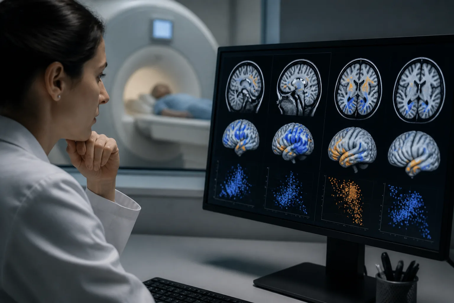
Researchers recently employed a method called Subtype and Stage Inference (SuStaIn) to analyze structural MRI data from the Autism Brain Imaging Data Exchange (ABIDE). This approach aims to identify subtypes of gray matter organization and track stage-like variations across individuals with ASD, moving beyond traditional group-level comparisons.
Two Consistent Gray Matter Subtypes Emerged
The analysis revealed two primary gray matter subtypes in ASD that remained consistent when considering both increased and decreased gray matter volumes relative to controls. The first subtype involved brain regions including the cerebellum, limbic system, and striatum. The second subtype was characterized by differences in the anterior frontal, cingulate, and insular cortices.
Clinical Associations
Exploratory analyses adjusting for covariates suggested that individuals in the first subtype with more advanced stage-like brain changes tended to have lower verbal intelligence quotient (IQ) scores. Meanwhile, those in the second subtype showed modestly higher scores on the Autism Diagnostic Observation Schedule (ADOS), a measure of autism symptom severity.
Structural Brain Network Differences
Further investigation into structural covariance networks—how brain regions co-vary structurally—revealed differences related to stage groups within each subtype. However, there was limited evidence for stable covariance differences between the two subtypes themselves.
Implications for Autism Research and Families
This study offers a new neuroanatomical framework to better understand the heterogeneity in ASD by identifying distinct gray matter patterns linked to different clinical features. Recognizing these subtypes may help tailor future research and interventions to more specific brain-behavior profiles within the autism spectrum.
Limitations and Future Directions
While the findings provide valuable insights, they are based on cross-sectional data and require further validation in longitudinal studies. Additionally, the clinical associations observed were modest and exploratory, underscoring the need for cautious interpretation.
For more detailed information, see the original research published in Brain Imaging and Behavior: source.
Download the images in this article