Study Reveals How Propionic Acid Alters Mitochondrial Function in Neuronal Cells

New Insights into Mitochondrial Changes Linked to Propionic Acid Exposure
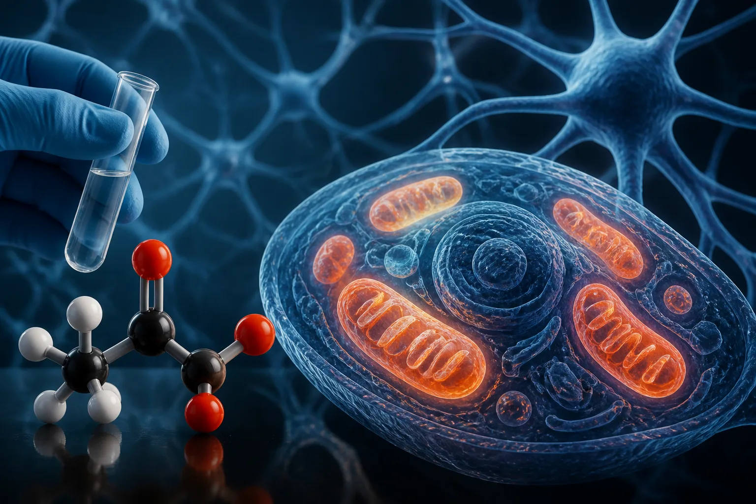
Recent research has shed light on how propionic acid (PPA), a compound often used in laboratory models to study neurological disorders such as Autism Spectrum Disorder (ASD), affects mitochondrial function in neuronal cells. The study focused on SH-SY5Y neuroblastoma cells, a commonly used model for human neurons, to explore how PPA exposure leads to changes in mitochondrial metabolism and structure.
Mitochondrial Dynamics Disrupted by Propionic Acid
The investigation revealed that PPA exposure significantly altered the expression of genes regulating mitochondrial dynamics, including those responsible for mitochondrial fusion and fission processes. Key proteins involved in these processes—L-OPA1 and MFN2 for fusion, DRP1 for fission—as well as LC3-II, a marker for mitochondrial quality control, were notably affected. These molecular changes were accompanied by increased occurrences of both fusion and fission events, as observed through confocal microscopy, indicating a remodeling of mitochondrial networks.
Impact on Mitochondrial Integrity and Energy Production
Alongside structural changes, PPA exposure impaired mitochondrial integrity and connectivity, disrupting the normal turnover of mitochondria within the cells. Functional assays demonstrated that these alterations led to deficits in cellular energy production, affecting both oxidative phosphorylation and glycolysis pathways. Further analysis of the respiratory chain showed a shift in the tricarboxylic acid (TCA) cycle flux, suggesting that mitochondrial substrate utilization was remodeled under PPA-induced stress.
Implications for Understanding Neurotoxicity and ASD Models
This study provides a detailed molecular signature of metabolic stress induced by PPA in neuronal cells, offering new perspectives on how mitochondrial dysfunction may contribute to neurotoxicity. While the findings are based on a cellular model and do not establish direct causation in human neurological conditions, they enhance understanding of mitochondrial involvement in complex disorders such as ASD.
Considerations for Families and Researchers
For families and individuals interested in the biological underpinnings of neurodevelopmental disorders, these findings highlight the importance of mitochondrial health in brain function. However, it is essential to recognize that this research is preliminary and conducted in vitro, meaning that further studies are necessary to determine how these cellular changes translate to clinical outcomes.
Study Limitations and Future Directions
The research was conducted using a neuroblastoma cell line, which, while informative, cannot fully replicate the complexity of human brain tissue. Additionally, the mechanisms by which PPA influences mitochondrial remodeling require further exploration to clarify their relevance in living organisms and their potential role in ASD pathology.
For more detailed information, the original study can be accessed here.
Download the images in this article