
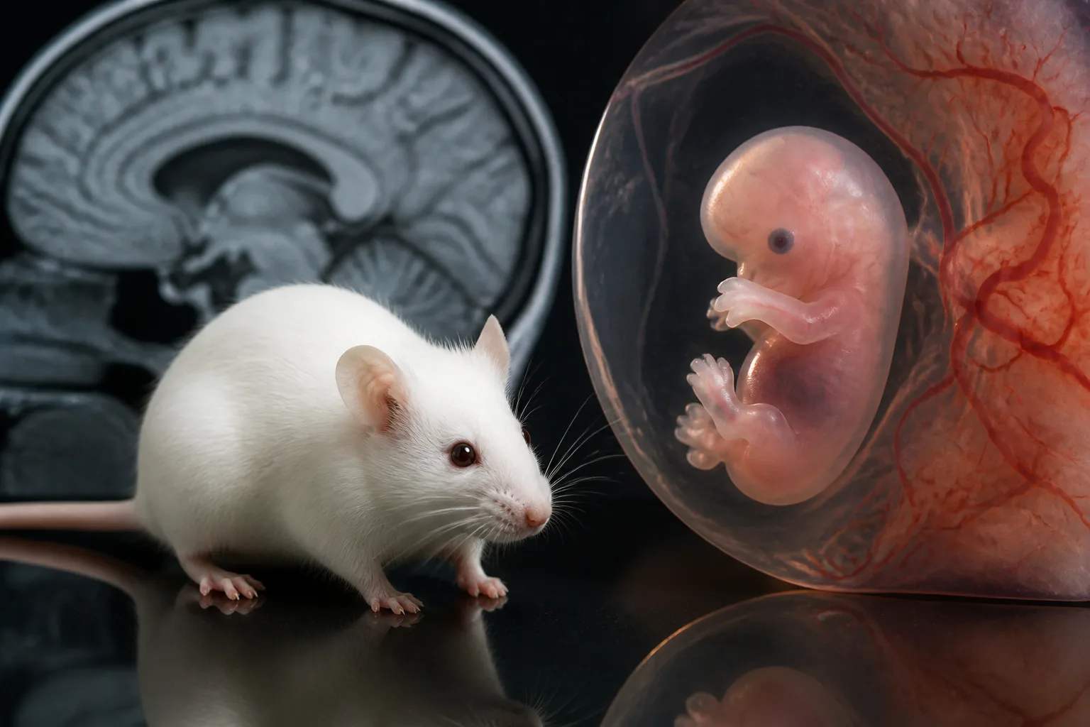
Prenatal Brain Development Differences in BTBR Mouse Model Linked to Neurodevelopmental Disorders

Study Explores Early Brain Development in BTBR Mice
The BTBR T+ Itpr3tf/J (BTBR) mouse strain, commonly used to study neurodevelopmental disorders, exhibits distinct brain anatomy abnormalities, including a complete absence of the corpus callosum. While adult BTBR brains have been extensively examined, less is known about how their brain development diverges prenatally from typical mouse strains.
Comparative Analysis of Fetal Brain Development
Researchers conducted a detailed histological comparison of fetal brain development between BTBR mice and the standard C57BL/6J (B6) strain. The focus was on the formation of the neocortex and midline brain structures at various embryonic stages.
Key Findings on Growth and Cortical Layers
At 12.5 days post coitum (dpc), BTBR fetuses weighed less than B6 controls but had similar somite counts, suggesting comparable overall developmental timing. By 15.5 dpc, BTBR mice showed a reduced proportion of the ventricular zone within the neocortex, although the total cortical thickness remained unchanged.
Midline Structural Alterations
Further examination revealed that midline cell populations positive for the glutamate aspartate transporter (GLAST) did not undergo the typical remodeling seen in B6 mice. This failure led to the absence of a GLAST-positive indusium griseum and a lack of callosal axon projections crossing the midline by 17.5 dpc in BTBR fetuses. In contrast, these features developed normally in B6 mice.
Implications for Understanding Neurodevelopmental Disorders
These prenatal differences in brain structure provide important insights into the early neuropathological changes associated with the BTBR mouse model. The absence of callosal connections and altered cortical layering may underlie some behavioral and neurological traits observed in this model.
Considerations and Context
While the BTBR strain offers valuable information about neurodevelopmental disorder mechanisms, these findings are specific to this mouse model and may not directly translate to human conditions. Further research is needed to clarify how these early brain alterations influence later functional outcomes.
For more detailed information, see the original study published in Biology: Fetal Cortical Layers and Midline Histological Alterations in the BTBR Mouse Model of Neurodevelopmental Disorders.
Download the images in this article