
New Study Finds Stable Individual Brain Connectivity Patterns but No Clear Autism Group Differences

Research Explores Brain Connectivity in Autism Spectrum Disorder
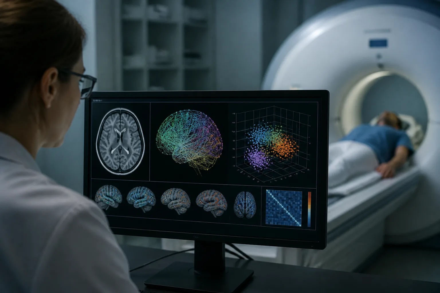
A recent study investigated whether differences in resting-state brain connectivity could reliably distinguish individuals with autism spectrum disorder (ASD) from neurotypical controls. Using an advanced geometric approach to analyze functional brain networks, researchers examined data from over 1,000 participants across multiple sites to assess the stability and diagnostic relevance of these connectivity patterns.
Key Findings: Individual Stability but No Group Differences
The study introduced a novel method that represents brain connectomes—maps of functional connections between brain regions—on a mathematical space called the symmetric positive-definite Riemannian manifold. This framework allowed quantification of how much an individual’s brain connectivity deviates from a normative model, termed “tangent-space dispersion.” Complementary analyses examined graph topology features.
Three main results emerged from the analysis of the Autism Brain Imaging Data Exchange (ABIDE I) dataset, which included 1,035 participants:
- Individual uniqueness and stability: The geometric descriptor of brain connectivity was highly reliable within individuals, showing consistent patterns across repeated measurements. After adjusting for differences related to the data acquisition site, the method could identify individuals within the same scanning session with 98.9% accuracy.
- No significant autism-related group differences: Statistical tests found no meaningful difference in the dispersion measure between ASD and control groups. Equivalence testing confirmed that any differences were negligible, with effect sizes near zero across various sample subsets. A minor effect seen in one analysis did not hold up under further scrutiny.
- Site effects dominate variance: Differences in data acquisition sites accounted for 69% of the variability in dispersion measures, while ASD diagnosis explained only 0.0001%. Moreover, connectivity deviations did not cluster into distinct subtypes nor predict symptom severity.
Implications for Autism Research and Brain Imaging
These findings suggest that while individual brain connectivity patterns are stable and uniquely identifiable, they do not serve as reliable biomarkers for ASD diagnosis or symptom severity. This may help explain why previous studies have reported inconsistent results regarding functional connectivity differences in autism.
The dominance of site-related variability highlights the challenges of multi-site neuroimaging studies and underscores the need for careful methodological controls when interpreting group differences.
Limitations and Considerations
Though the study employed rigorous methods and a large, multi-site dataset, it focused on resting-state functional MRI data and a specific geometric framework. The absence of group differences does not rule out other brain features or modalities that might relate to ASD. Additionally, the findings emphasize correlation rather than causation, and further research is needed to explore the complex neurobiology of autism.
For more detailed information, the original research article is available at PubMed.
Download the images in this article